Answering Dr. Robert Mendelsohn's X-ray Questions
Dr. Robert Mendelsohn, M.D. wrote an excellent book in 1981 about modern medicine’s negative and biased approach towards women’s health. The book is called “Male Practice: How Doctors Manipulate Women.” I recommended one read it today for its prophetic words and applicable nature in our current healthcare situation.
In the book, there is an entire chapter dedicated to the explanation of medicine’s unnecessary X-ray overuse and the ill-effects they bring. Dr. Mendelsohn calls X-rays “the most dangerous weapons in Modern Medicine’s arsenal.” At the end of the chapter, he provides a list of 9 questions that every patient should ask their doctor on the validity of taking an X-ray.
As a Chiropractor who takes X-rays, I found the chapter challenging, enlightening, and most importantly, a teachable moment. Addressing the questions brought up in the chapter head-on not only will help validate the use of taking X-rays from a chiropractic standpoint but will only help further delineate and differentiate between why a chiropractor would take X-rays versus why a medical doctor would take X-rays. Even though both chiropractic and medicine utilize X-ray imaging in their practice, the reasoning for taking the X-ray and the application of the X-ray are completely different.
The difference in application boils down to two similar but different words: diagnosis and analysis. The main reason (but not the only reason) a chiropractor would take an X-ray is to assist in the analysis of a vertebral subluxation. The main reason (and only reason) a medical doctor would take an X-ray is to assist in the process of differential diagnosis.
I would like to now answer those 9 questions proposed by Dr. Mendelsohn. I’ve altered some of the lexicon of the questions to better fit the Chiropractic vocabulary, but the sentiment is the same. As a patient, you should ask these questions regardless if you are in the presence of a chiropractor or medical doctor and they want to take X-rays.
What are you looking for?
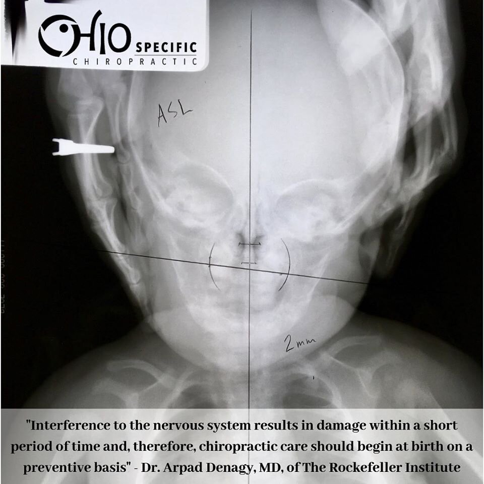
As a chiropractor, the main reason I am taking X-rays and what I am looking for from the X-rays is a vertebral subluxation. A vertebral subluxation is a spinal bone that has lost its normal position to an extent less than a dislocation and has occluded a foramen. It also has to be putting pressure upon nerves and interfering with the transmission of a mental impulse.
X-rays, or Spinographs (a chiropractic term), help me answer the first two parts to the definition: Has the vertebra lost normal position to an extent less than a dislocation and has that vertebra occluded a foramen. Before answering the first two parts of the subluxation definition, I will actually answer the last two parts first: Is there pressure upon nerves and is there interference with the transmission of a mental impulse.
By first testing if the neurological components are present, I have a higher degree of certainty that taking X-ray images will confirm the osseous components. If the neurological tests do not indicate nerve pressure and mental impulse interference, then X-rays will not be taken at that time. This approach cuts down on unnecessary X-rays.
What are the chances that you’ll find it if you take these X-rays?
While not 100% certain, the spinographs provide the Chiropractor a great opportunity to locate the osseous component of the subluxation. Clinically, if a subluxation is what we say it is, then 100% of the time the occlusion and malposition will be present in the osseous structures and be found on the X-rays.
A large portion of the ability to “find” the subluxation though, will be up to the competency of the Chiropractor to analyze the X-rays to determine the exact nature of the bony component of the subluxation. To reiterate, I will perform the non-invasive neurological analysis before even considering taking X-rays to increase the probability that a subluxation is present and the osseous component is found on the X-ray.
Can you find what you are looking for by some less hazardous means?
Not at all with the same precision. Non-imaging analysis such as palpation, leg checks and symptoms cause too many variables and not enough constants; both from the patient and the doctor. Subjective interpretation plagues all analysis, but even more so in things such as palpation and symptoms.
Experiments performed early in the development of Chiropractic proved that using palpation and symptoms alone had serious error outcomes in determining the subluxated vertebra. The initial tests showed that up to 83% of the population has malformations in their osseous structures. This results in asymmetry of spinous and transverse processes; the bony projections used in palpation.
With this newfound information, experiments were performed that used palpation and symptoms to analyze subluxations. In non-hyperbolic fashion, the results found 0% reliability with regards to objective subluxation analysis. Many Chiropractors today still just rely on palpation and symptoms for their subluxation analysis which should send up a red flag to a concerned patient. Objective imaging provides a snapshot of something that can’t be visualized, cutting down on potential hazardous outcomes.
If you find it, is it something you can correct?
The short answer is ‘Yes’. The correction of the subluxation is in the hands of both the competent chiropractor and committed patient. The Chiropractor applies the adjustic force while the patient adapts the force into a correction.
Chiropractic is a joint effort between the doctor and patient. Chiropractors spend a minimum of 3 ½ years in school learning proper analysis and technique in the adjustment of the subluxation. As a chiropractic student, I was required to adjust 50 fellow students, and 200 outpatients in the clinic.
Along with reaching my required adjustment numbers through the curriculum, I also took elective classes to further hone my adjustive skills. Even further beyond that, I took part in Chiropractic mission trips to Nicaragua where I had the opportunity to adjust over 2,000 children and adults. My educational and clinical experience gives me confidence in obtaining the goal of the adjustment.
Does the careplan entail any risks? If not, why can’t you just assume I have the condition, skip the X-ray, give me that care, and see if it works?
Yes, there are risks involved with taking X-rays. Contextually, all healthcare procedures caring risk, no matter what they are. Furthermore, there are also risks involved with not taking X-rays. From a Chiropractic understanding, the risk of negative outcomes increase if we do not know exactly how the bones misalign.
To guess at which vectors and angles our specific adjustic force needs to be applied can lead to undesirable consequences. For example, let’s say you have a simple fracture of your femur. A simple fracture is a type of fracture that does not break the skin. And since it does not break the skin, it can be hard to determine the exact direction and degree of the fracture. Would you allow the orthopedic surgeon to set the bone without knowing the specific direction and degree at which the bone fractured? The orthopedic doctor could assume that the bone fractured 20 millimeters to the right and at 10 degrees superior, but should you take their assumption without knowing precisely before they set the bone?
Assumptions should not be the bedrock of any healthcare application. There is an old maxim in Chiropractic that states: “Why Chiropractic X-rays? To see is to know, not to see is to guess, and we won’t guess about your health."
When was the last time your X-ray equipment was checked for safety?
In the state of Ohio, the Department of Health checks our X-ray equipment every 3 years through a series of safety protocol inspections. Quality assurance items are reviewed and X-ray equipment is tested. The performance inspection includes the following: Current certificate of registration, Regulations applicable to facility type, Prior violations and corrective action review, Notice-to-Employees postings, Operator training, Personnel monitoring, Radiologic licenses, Preventative maintenance, Quality control tests: daily, monthly, quarterly, Area radiation surveys, X-ray equipment inventory, X-ray equipment safe operating procedures, X-ray equipment receipts, transfers and disposals, Fluoroscopic equipment: tests by the medical physicist, Computed tomography equipment: tests by the medical physicist, Radiation therapy equipment: tests by the medical physicist, Any self-referral approval letters, Any Food & Drug Administration (FDA) X-ray equipment variances, Exposure, kVp and timer reproducibility, kVp and timer accuracy, Half-value layer, Exposure switch position and function, Entrance-skin exposure for examination, Collimator and light field congruency, Collimator beam size an alignment factors, Safety interlocks
Will it be operated by a trained technician who knows what he or she is doing and will keep the radiation dosage to a minimum?
Chiropractors, in general, have over 300 hours of radiology education, including diagnosis, equipment use and patient placement. The study of radiology accounts for 12% of our clinical training in college. I pride myself on taking my own X-rays. It is important for me to be with a new patient every step of the way on the initial exam.
In general, medically-trained X-ray technicians do not learn the small intricate details needed to take a proper Chiropractic upper cervical specific X-ray series. Exact placement is integral for upper cervical specific films. Since I will be the one analyzing the films and making my adjustment based on my analysis, it is my job and your right as a patient to demand the highest quality of care.
Based on your age, gender and weight I modify the radiation voltage to prevent needless radiation dosage.
What form of shielding or other protection will you furnish me?
Lead aprons are provided and cover areas that are not necessary for the X-ray viewing series. Lead is often used in protection shields because it helps prevent the scattering of radiation. Lead is a heavy metal and is very dense in nature. The high density means the atoms that comprise lead are packed tightly together. Compared to other metals such as copper and iron, lead has a higher density and atomic number. The density level prevents gamma rays and X-rays from passing through them.
Lead aprons usually cover the reproductive organs of both males and females, especially if an X-ray is taken in the lumbar and/or thoracic region. The majority of my X-rays are taken of the upper cervical spine, so radiation scatter to these regions of the body are very rare. The major concern with taking cervical X-rays is the proximity of the thyroid gland to this area.
The thyroid gland is involved in many metabolic and hormonal processes; a malfunctioning thyroid can lead to a variety of ill-conditions. The major difference between an upper cervical specific X-ray series and a regular cervical series is the location of the central ray. The central ray is the point of the tube where the radiation is created. From this point, the X-rays diverge out from the tube in a cone-shaped projection. The central ray also has the greatest concentration of radiation, lessening as it diverges out.
With a regular cervical series, the central ray is located at the C5 vertebra, just posterior to the thyroid gland. With an upper cervical series, the central ray is located at the C1 vertebra. This means there is a higher concentration of radiation to the thyroid gland with a regular cervical series than with an upper cervical series. Although the radiation risk is low in either series, exposure is less in the upper cervical series.
What dose of radiation will I receive?
Radiation exposure is a warranted health concern of the public and should not be glossed over by healthcare professionals as something inert. I do think it is important though to put perspective into the conversation when it comes to dosage. When talking with my patients, they are always surprised to find out that the general population receives natural radiation all the time, and usually from sources not advertised as producing radiation.
The average person in the United States receives an effective dose of about 3.1 mSV per year coming from natural radiation and cosmic radiation. The predominant source of natural radiation is found in the air we breathe, inhaled as radon. The other main sources are found in our food and water, ingested as uranium, thorium, and radium.
Radiation exposure has increased over the last 20 years though, with the majority of the increase coming from healthcare sources. The major healthcare source of exposure comes from X-rays. With all the negative press that standard X-ray imaging receives, other imaging procedures seem to skid by unnoticed with regards to radiation dosage.
One imaging procedure that seems to get a “hall pass” in the radiation debate is CT Scans. CT Scans are a series of X-ray images that are taken from different angles to create cross-sectional sections of the body. One CT scan can total an average of 6 mSv, double the normal amount of radiation a person is exposed to in an entire year.
As an Upper Cervical Chiropractor, I take a 3 View Cervical Series. With this series of X-rays, the total dose of radiation averages out to be around 0.6 millisievert (mSv). This is using an 18-year-old person as a point of reference.
These 9 questions and answers are not exhaustive when it comes to X-ray validity and patient concerns. I hope though that they at least answer some inquiries when it comes to X-rays. This is a place to start in the conversation and hopefully eases the patient’s reservations about starting Chiropractic care.
- Jarek Esarco, D.C.
Related Blogs:
100+ Years of Pediatric Spinography
Adjusting Children: A New Age Niche or an Established Institute Foundation?